Best Digital X-ray Centre in Meerut

Home Radiology Services Digital X-ray
Best Digital X-ray Centre in Meerut

Introduction

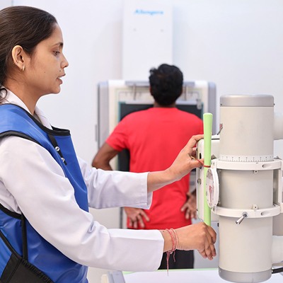
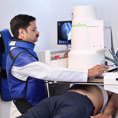
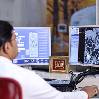
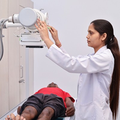
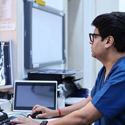
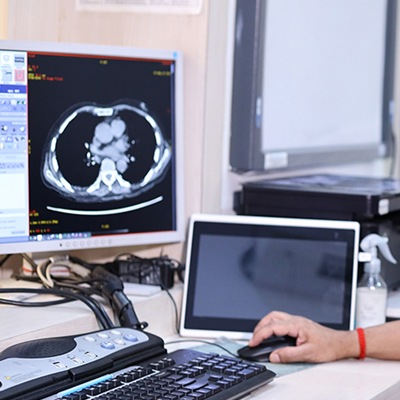
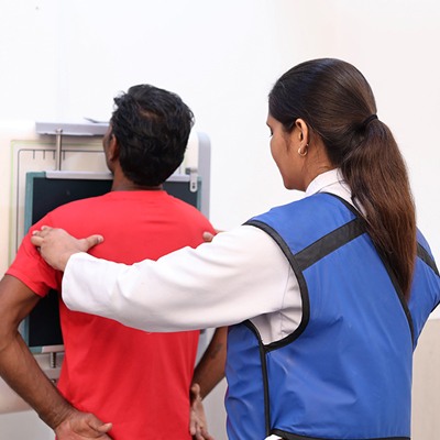
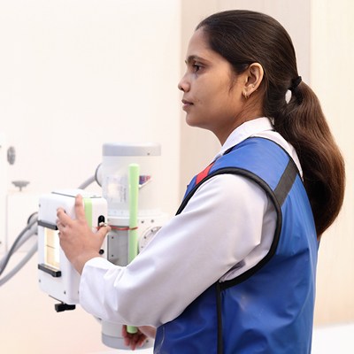
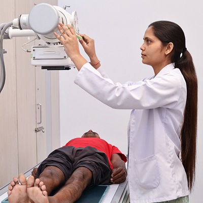
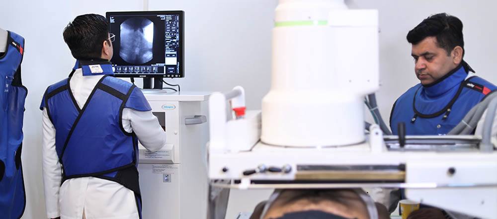
Digital X-ray is an advanced imaging technique that utilizes X-rays to produce clear, detailed images of bones, organs, and tissues. At Tesla Imaging & Diagnostic Centre, Meerut, under the expert guidance of Dr. Shalabh Bansal, we employ state-of-the-art digital X-ray technology to ensure accurate and efficient diagnosis. Compared to traditional film-based X-rays, digital X-rays offer several advantages, including faster image acquisition, improved image quality, and the ability to manipulate and enhance images for better visualization.

Chest X-Ray:

A Chest X-ray is a common imaging technique used to examine the lungs, heart, and blood vessels. At Tesla Imaging & Diagnostic Centre, Meerut, under the expert guidance of Dr. Shalabh Bansal, chest X-rays are performed to diagnose conditions such as pneumonia, pneumothorax, heart failure, and lung cancer.

Chest X-Ray Centre in Meerut
KUB & Abdomen Centre in Meerut

KUB & Abdomen:

A KUB (Kidneys, Ureters, Bladder) and Abdomen X-ray provides images of the abdominal organs, including the kidneys, ureters, bladder, liver, spleen, and intestines. At Tesla Imaging & Diagnostic Centre, Meerut, under the care of Dr. Shalabh Bansal, KUB X-rays are used to evaluate abdominal pain, kidney stones, and other abdominal conditions.

Barium Meal Follow Through (BMFT):

A Barium Meal Follow Through is a diagnostic procedure that involves drinking a liquid containing barium sulfate, a contrast material. At Tesla Imaging & Diagnostic Centre, Meerut, under the guidance of Dr. Shalabh Bansal, a series of X-rays are taken to track the barium's movement through the digestive system, helping to diagnose conditions like ulcers, tumors, and inflammatory bowel disease.

Barium Meal Follow Through (BMFT) Centre in Meerut
Barium Enema Centre in Meerut

Barium Enema:

A Barium Enema involves introducing a barium solution into the rectum to visualize the large intestine. At Tesla Imaging & Diagnostic Centre, Meerut, under the expertise of Dr. Shalabh Bansal, this procedure helps diagnose conditions such as colitis, diverticulitis, and colorectal cancer.

IVP - Intra Venous Pyelography / Urography:

An IVP (Intra Venous Pyelography) or Urography involves injecting a contrast dye into a vein, allowing for visualization of the kidneys, ureters, and bladder. At Tesla Imaging & Diagnostic Centre, Meerut, under the care of Dr. Shalabh Bansal, IVP is used to diagnose kidney stones, kidney infections, and other urinary tract conditions.

Intra Venous Pyelography / Urography Centre in Meerut
Hystero Salpingography Centre in Meerut

HSG - Hystero Salpingography:

A Hysterosalpingography (HSG) is an X-ray examination of the uterus and fallopian tubes. At Tesla Imaging & Diagnostic Centre, Meerut, under the guidance of Dr. Shalabh Bansal, HSG involves injecting a contrast dye into the uterus to assess the fallopian tubes for blockages or abnormalities, which can affect fertility.

Sono Salpingography:

Sono Salpingography is an ultrasound-guided procedure to assess the fallopian tubes. At Tesla Imaging & Diagnostic Centre, Meerut, under the expertise of Dr. Shalabh Bansal, this technique involves injecting a saline solution into the uterus to visualize the fallopian tubes and identify any blockages.

Sono Salpingography Centre in Meerut
Sinogram Centre in Meerut

Sinogram:

A Sinogram is a type of CT scan specifically designed to image the paranasal sinuses. At Tesla Imaging & Diagnostic Centre, Meerut, under the care of Dr. Shalabh Bansal, sinograms are used to diagnose sinus infections, polyps, and other sinus-related conditions.

T-tube Cholangiogram:

A T-tube Cholangiogram is an X-ray examination of the bile ducts. It is performed on patients who have undergone biliary surgery and have a T-tube in place. At Tesla Imaging & Diagnostic Centre, Meerut, under the guidance of Dr. Shalabh Bansal, this procedure helps assess bile duct patency and identify any complications.

T-tube Cholangiogram Centre in Meerut
Retrograde Urography Centre in Meerut

RGU - Retrograde Urography:

Retrograde Urography is a diagnostic procedure involving the insertion of a catheter into the ureters to inject contrast dye directly into the kidneys. At Tesla Imaging & Diagnostic Centre, Meerut, under the expertise of Dr. Shalabh Bansal, RGU is used to visualize the urinary tract when conventional imaging methods are not sufficient.

MCU - Micturiting Cysto Uretherogram:

A Micturiting Cysto Uretherogram (MCU) is an X-ray examination of the bladder and urethra during urination. At Tesla Imaging & Diagnostic Centre, Meerut, under the care of Dr. Shalabh Bansal, MCU is used to diagnose conditions like vesicoureteral reflux, bladder outlet obstruction, and other urinary tract abnormalities.

Micturiting Cysto Uretherogram Centre in Meerut

Faqs

Digital X-ray FAQs at Tesla Imaging & Diagnostic Centre, Meerut

The primary difference between Digital X-rays and traditional X-rays is the way the images are captured and processed. Digital X-rays use digital sensors to capture images, which are then stored electronically, offering quicker results, better image quality, and reduced radiation exposure compared to traditional film-based X-rays.

Digital X-rays expose patients to significantly less radiation than traditional X-rays. The exact amount of radiation depends on the type of X-ray being performed, but Digital X-rays generally use up to 90% less radiation, making them a safer option for diagnostic imaging.

Digital X-rays are faster because the images are captured electronically and can be viewed on a computer screen almost immediately. This eliminates the need for film development, allowing healthcare providers to analyze the images and diagnose conditions much more quickly.

Yes, Digital X-rays often provide clearer and more detailed images than traditional X-rays. The ability to enhance and zoom in on digital images allows healthcare providers to detect smaller abnormalities and diagnose conditions more accurately, making them a superior diagnostic tool.

You may experience slight tenderness or soreness in your breasts for a few days after the mammogram.