Types of Ultrasound
There are several types of ultrasound scans, each designed to examine specific parts of the body. Some common types include:
Trust 18+ years of diagnostic experience in Meerut. Call Us Today:
Call Now: +91-9456902244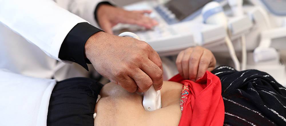
There are several types of ultrasound scans, each designed to examine specific parts of the body. Some common types include:
Trust 18+ years of diagnostic experience in Meerut. Call Us Today:
Call Now: +91-94569022443D/4D Sonography offers a detailed, three-dimensional or even real-time view of the fetus, providing parents with a remarkable experience. At Tesla Imaging & Diagnostic Centre, Meerut, under the expertise of Dr. Shalabh Bansal, this advanced technology allows for a more comprehensive assessment of fetal development and well-being.
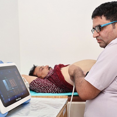
FibroScan, also known as Elastography, is a non-invasive technique used to assess liver stiffness. Offered at Tesla Imaging & Diagnostic Centre, Meerut, under the supervision of Dr. Shalabh Bansal, FibroScan helps in detecting liver fibrosis and evaluating liver health without the need for a liver biopsy.
Orbital or Ocular Ultrasound provides detailed images of the eye and its surrounding structures. At Tesla Imaging & Diagnostic Centre, Meerut, this ultrasound is used to diagnose conditions like glaucoma, cataracts, retinal detachments, and eye tumors.
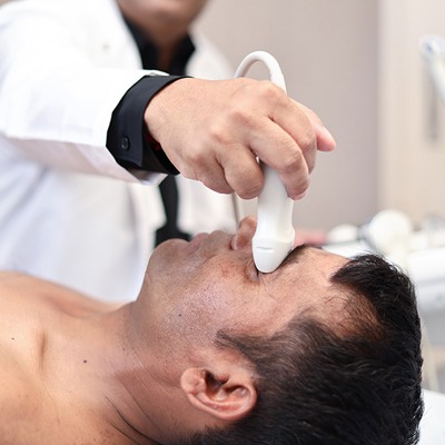
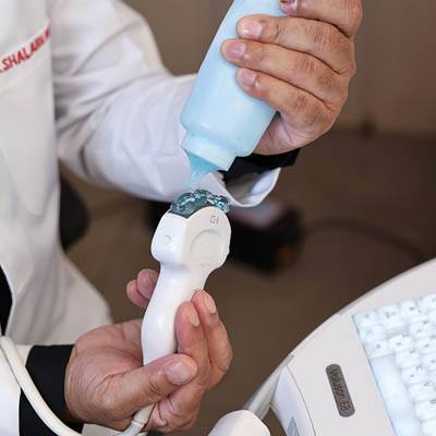
Sialography involves injecting contrast dye into the salivary glands to visualize their ducts and structures. Performed at Tesla Imaging & Diagnostic Centre, Meerut, under the guidance of Dr. Shalabh Bansal, this procedure helps diagnose salivary gland conditions like stones, inflammation, and strictures.
Sono Mammography, or breast ultrasound, is a non-invasive imaging technique used to examine breast tissue. At Tesla Imaging & Diagnostic Centre, Meerut, under the care of Dr. Shalabh Bansal, breast ultrasound is often used as a complementary diagnostic tool alongside mammograms to assess breast abnormalities.
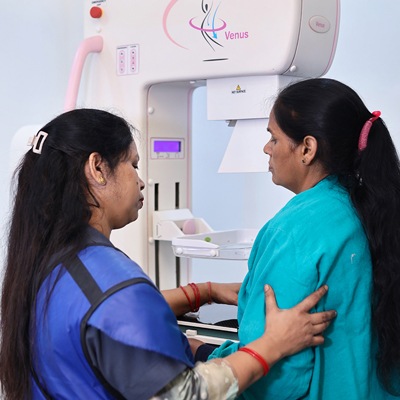
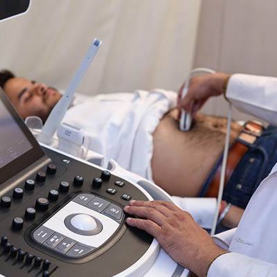
Scrotal Ultrasound provides images of the testicles and surrounding structures. Performed at Tesla Imaging & Diagnostic Centre, Meerut, under the guidance of Dr. Shalabh Bansal, this ultrasound helps diagnose conditions like testicular torsion, epididymitis, and testicular masses.
Shoulder Ultrasound is used to evaluate the soft tissues, tendons, and muscles of the shoulder joint. At Tesla Imaging & Diagnostic Centre, Meerut, under the expertise of Dr. Shalabh Bansal, this ultrasound helps diagnose rotator cuff tears, bursitis, and other shoulder problems.
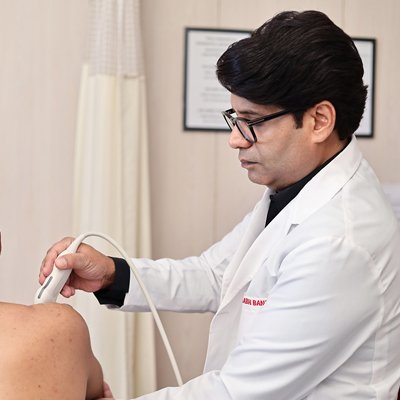
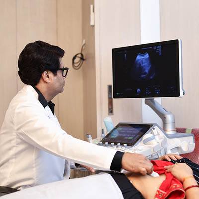
Transvaginal Scan (TVS) is a type of ultrasound performed by inserting a probe into the vagina. At Tesla Imaging & Diagnostic Centre, Meerut, under the care of Dr. Shalabh Bansal, 3D/4D TVS provides detailed images of the pelvic organs, including the uterus, ovaries, and fallopian tubes.
Follicular Study and Antral Follicular Count (AFC) are ultrasound-based assessments of ovarian function. Performed at Tesla Imaging & Diagnostic Centre, Meerut, under the guidance of Dr. Shalabh Bansal, these studies help evaluate fertility and monitor ovulation.
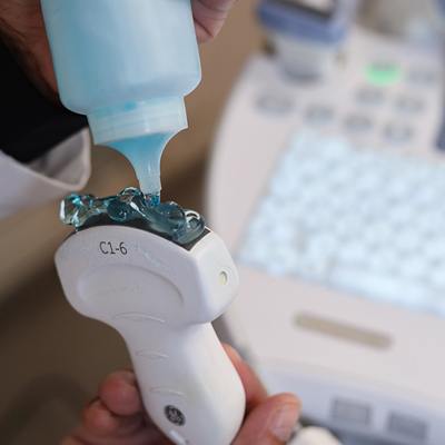
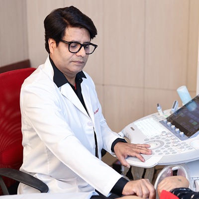
Prostate Ultrasound, or Transrectal Ultrasound (TRUS), is used to examine the prostate gland. At Tesla Imaging & Diagnostic Centre, Meerut, under the expertise of Dr. Shalabh Bansal, TRUS is used for diagnosing prostate cancer, prostatitis, and benign prostatic hyperplasia (BPH).
Ultrasound-Guided Fine Needle Aspiration and Biopsy (USG-FNAC/Biopsy) is a minimally invasive procedure performed at Tesla Imaging & Diagnostic Centre, Meerut under the guidance of Dr. Shalabh Bansal. Ultrasound is used to precisely guide the needle for obtaining tissue samples for diagnosis.
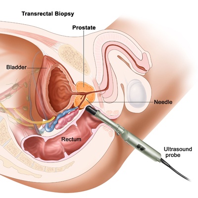
TRUS-guided Prostatic Biopsy is a procedure to obtain tissue samples from the prostate gland. At Tesla Imaging & Diagnostic Centre, Meerut, under the expertise of Dr. Shalabh Bansal, ultrasound is used to guide the biopsy needle for accurate sampling.
Renal Doppler Ultrasound evaluates blood flow to the kidneys. Performed at Tesla Imaging & Diagnostic Centre, Meerut, under the guidance of Dr. Shalabh Bansal, this ultrasound helps diagnose kidney artery stenosis, kidney transplants, and other kidney-related conditions.
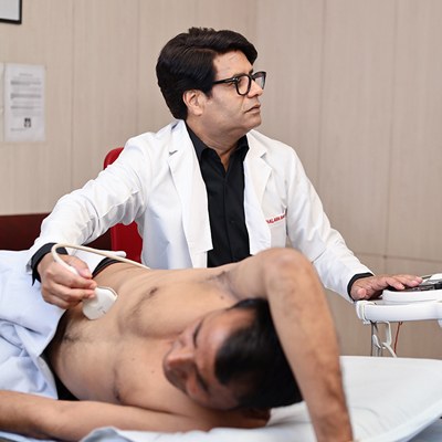
Portal Doppler Ultrasound assesses blood flow in the portal vein, which carries blood from the intestines to the liver. Performed at Tesla Imaging & Diagnostic Centre, Meerut, under the expertise of Dr. Shalabh Bansal, this ultrasound helps diagnose liver diseases and portal hypertension.
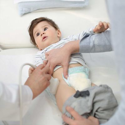
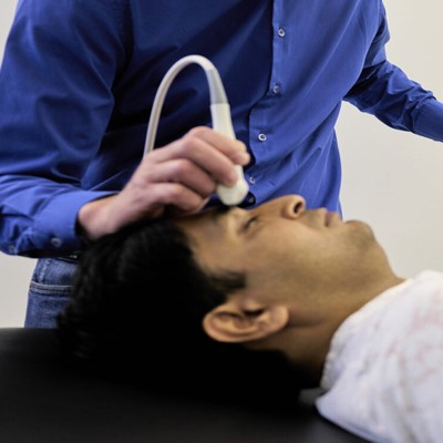
Brain Ultrasound, primarily used in infants, is a non-invasive technique to evaluate the brain. At Tesla Imaging & Diagnostic Centre, Meerut, under the care of Dr. Shalabh Bansal, brain ultrasound helps diagnose conditions like brain bleeds, hydrocephalus, and brain infections.
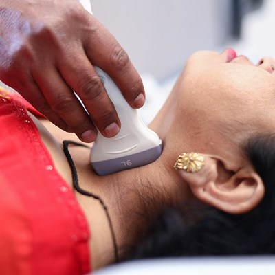
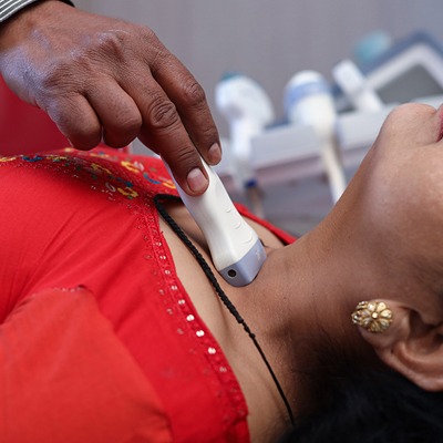
Neck Ultrasound examines the thyroid gland, lymph nodes, and other neck structures. Performed at Tesla Imaging & Diagnostic Centre, Meerut, under the guidance of Dr. Shalabh Bansal, this ultrasound helps diagnose thyroid disorders, lymph node enlargement, and other neck-related conditions.
Thyroid and Parotid Ultrasound focuses on imaging the thyroid and parotid glands. Performed at Tesla Imaging & Diagnostic Centre, Meerut, under the expertise of Dr. Shalabh Bansal, this ultrasound helps diagnose thyroid nodules, thyroiditis, and parotid gland inflammation.
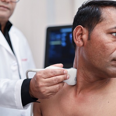
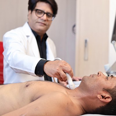
Carotid Doppler Ultrasound assesses blood flow in the carotid arteries, which supply blood to the brain. Performed at Tesla Imaging & Diagnostic Centre, Meerut, under the guidance of Dr. Shalabh Bansal, this ultrasound helps detect carotid artery stenosis and reduce the risk of stroke.
Chest Ultrasound provides images of the lungs, heart, and chest wall. Performed at Tesla Imaging & Diagnostic Centre, Meerut, under the care of Dr. Shalabh Bansal, this ultrasound is used to diagnose pleural effusions, pneumothorax, and other chest conditions.
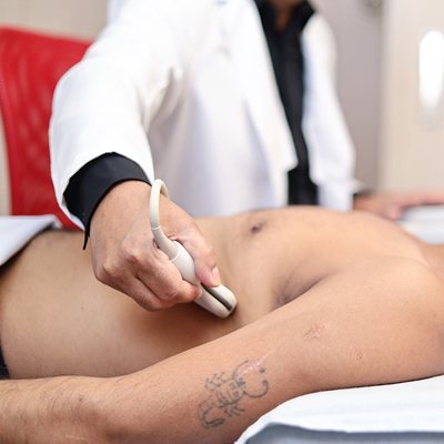
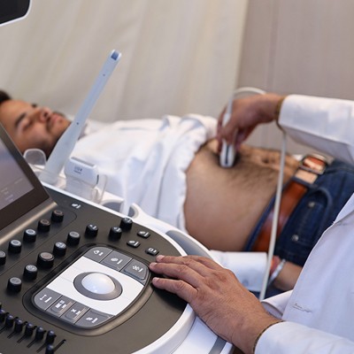
Abdominal Ultrasound examines organs like the liver, gallbladder, pancreas, spleen, kidneys, and bladder. Performed at Tesla Imaging & Diagnostic Centre, Meerut, under the guidance of Dr. Shalabh Bansal, this ultrasound helps diagnose various abdominal conditions.
Penile Ultrasound is used to evaluate the penis and its blood flow. Performed at Tesla Imaging & Diagnostic Centre, Meerut, under the expertise of Dr. Shalabh Bansal, this ultrasound helps diagnose erectile dysfunction and Peyronie's disease.
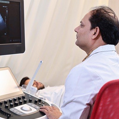
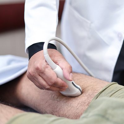
Peripheral Vascular Doppler Ultrasound assesses blood flow in the arteries and veins of the arms and legs. Performed at Tesla Imaging & Diagnostic Centre, Meerut, under the guidance of Dr. Shalabh Bansal, this ultrasound helps diagnose peripheral artery disease, deep vein thrombosis, and other vascular conditions.
Pediatric Hip Ultrasound is used to evaluate hip development in infants. Performed at Tesla Imaging & Diagnostic Centre, Meerut, under the care of Dr. Shalabh Bansal, this ultrasound helps diagnose developmental dysplasia of the hip (DDH).
